Upload a chest film, wait a few seconds, and get back a list of suspected findings with the abnormal regions outlined in colour. That is AI X-ray analysis as most people meet it for the first time, and the demo is genuinely impressive.
It is also the part that gets misread. The model is not reading the film the way a radiologist reads it, and it is not making a diagnosis. It is scoring probabilities and showing you which pixels moved those probabilities. Understanding that difference is the whole game — it decides whether the tool saves your team hours or quietly creates risk.
Here is the full pipeline, pixels in to explanation out, plus the four places it breaks.
What happens between upload and result
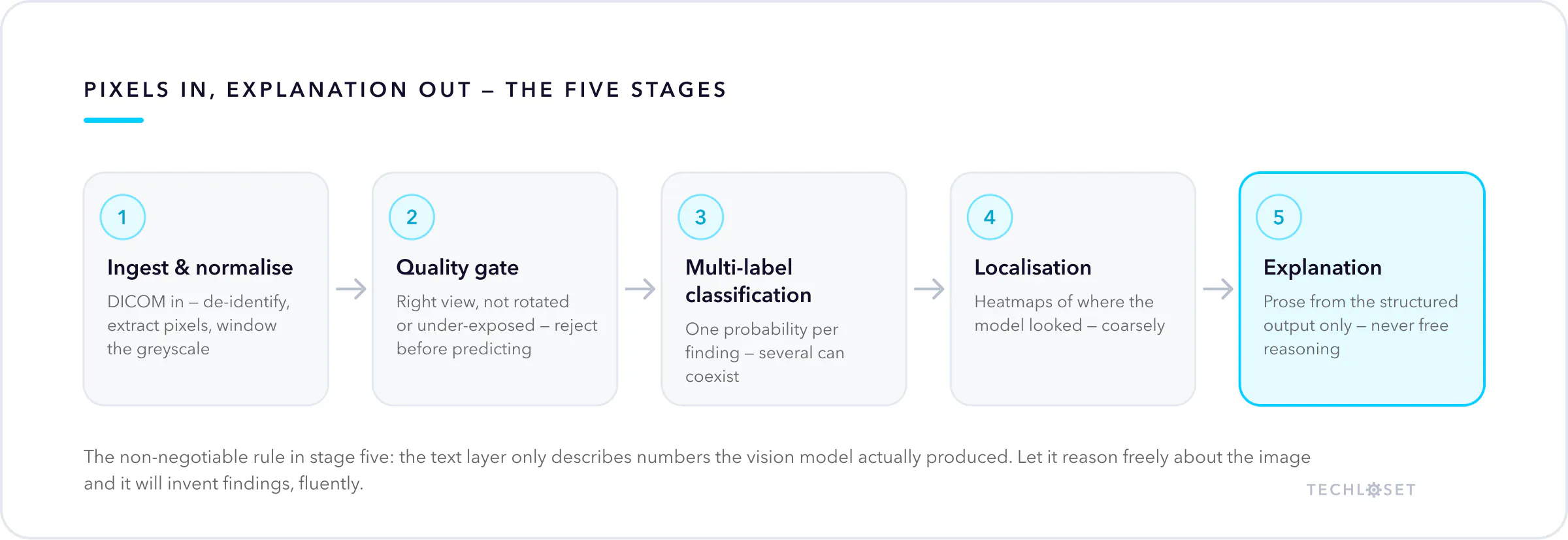
1. Ingest and normalise. Clinical images arrive as DICOM, not JPEG — a wrapper carrying the pixel data plus patient identifiers, study metadata, and acquisition settings. The file is de-identified, the pixel array is extracted, and windowing is applied so the greyscale range the model sees is consistent. Two X-rays of the same chest from two machines can differ more in exposure than two different patients do on one machine, so this step matters more than it sounds.
2. Quality gate. Before any prediction, a sane system checks it received what it expected. Is this a frontal chest film or a lateral knee? Is it rotated, clipped, or under-exposed past usefulness? A model asked to grade an image outside its training distribution will still return a confident number, which is exactly the failure you do not want reaching a clinician.
3. Multi-label classification. The core model — typically a convolutional network or vision transformer — outputs an independent probability for each of a fixed set of findings. "Multi-label" is the important word: a film can show consolidation and an effusion and an enlarged heart at once, so these are not competing options in a single choice. Public benchmarks like ChestX-ray14 and CheXpert established the ~14-finding convention most tools still follow.
4. Localisation. Saliency methods such as Grad-CAM work backwards from the prediction to the regions that drove it, producing the heatmap overlay you see in demos. This is genuinely useful and genuinely coarse — it shows where the model looked, which is not always the same as where the finding is.
5. Explanation and reporting. A language model turns the structured output — finding, probability, region — into readable prose and answers follow-up questions about it. The non-negotiable engineering rule: the text layer must only describe numbers the vision model actually produced. Let it reason freely about the image and it will invent findings fluently.
The four limits nobody puts in the demo
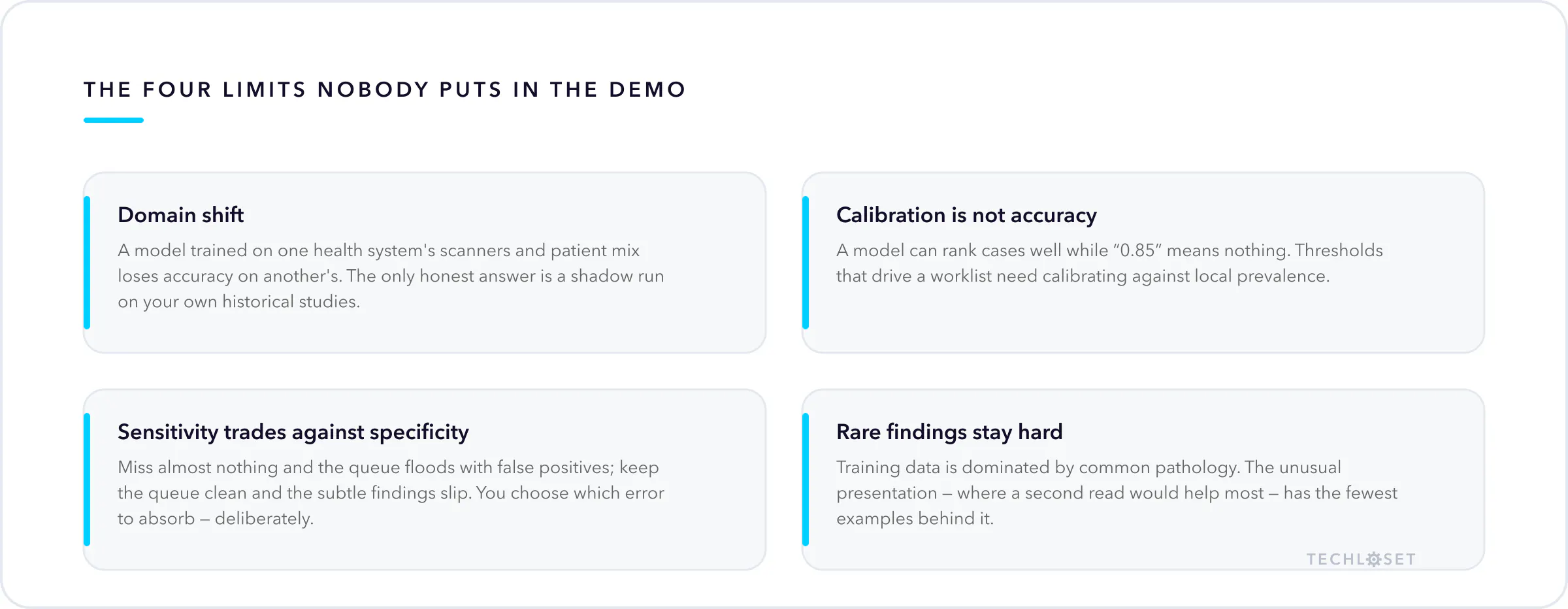
Domain shift is the big one. A model trained on one health system's scanners, positioning habits, and patient mix loses accuracy on another's. Performance on the vendor's held-out test set tells you little about performance on your equipment. The only honest answer is a shadow run on your own historical studies before anyone trusts it live.
Calibration is not accuracy. A model can rank cases well while its raw probabilities are meaningless — "0.85" may not mean 85% of such cases are positive. If a threshold is going to drive a worklist or an alert, the scores behind it need calibrating against local prevalence, not accepting at face value.
Sensitivity and specificity trade against each other, always. Tuned to miss almost nothing, a model floods the queue with false positives and staff learn to dismiss it. Tuned for a clean queue, it misses the subtle findings that were the entire reason to buy it. There is no setting that escapes this; there is only choosing which error you would rather absorb, deliberately.
Rare findings stay hard. Training data is dominated by common pathology. The unusual presentation — the one where a second read would genuinely help — is the one with the fewest examples behind it.
Assistive, not diagnostic — and why the wording matters
An AI X-ray tool flags, ranks, highlights, and drafts. A clinician interprets, decides, and signs. That boundary is not marketing caution, it is regulatory reality: many cleared radiology tools in the US are authorised for computer-aided triage — reordering the worklist so time-critical studies surface first — rather than for rendering a diagnosis. In the EU, the AI Act places medical AI in its high-risk tier, with obligations attached.
So the honest, defensible claim is narrow and still valuable: the urgent film gets looked at sooner, and the second pair of eyes never gets tired. Any vendor selling you more than that is either uncleared or overstating, and both are your problem after the sale.
The same rule applies to educational and pre-visit tools built on this stack — including the X-ray analysis assistant we build. Analysis and explanation, with a human making every call that matters.
What it is actually good for
Strip out the overclaiming and a real set of jobs remains:
- Worklist triage. Surfacing likely-critical studies first, so turnaround time on the cases that matter drops without hiring.
- Second-read backup. Catching the incidental finding at the end of a long shift, when attention is thinnest.
- Follow-up capture. Flagging studies whose recommended follow-up was never booked — a well-documented leak in almost every imaging operation, and pure recovered revenue when closed.
- Report drafting. Producing the structured first pass a radiologist edits instead of dictates.
- Teaching. Letting trainees ask "why this region?" against a labelled archive, safely, with no patient waiting on the answer.
Note what those have in common: each one saves a scarce human's minutes, or stops something from being dropped. None of them replaces the read.
FAQ
Is AI X-ray analysis accurate enough to use clinically? On common findings, published models perform in the range of trained readers on the datasets they were built for. That is not the same as performing that way on your images — validate locally before deploying, and keep a clinician in the loop regardless.
Can it replace a radiologist? No. Current tools output probabilities and regions; they do not integrate history, priors, symptoms, and clinical judgement into a signed interpretation. Their value is in triage and second-read support.
What about patient data? X-rays are PHI, including the DICOM metadata. HIPAA applies end to end: de-identification before processing, encryption in transit and at rest, audit logging, a signed BAA with any vendor touching the pixels.
How much data do you need to build one? Less than most people assume, if you transfer-learn from a pretrained backbone — thousands, not millions, of labelled studies. Label quality dominates label quantity. We break the build down here.
The short version
AI X-ray analysis is a probability engine with a highlighter, not a radiologist in a box. Judged as the second thing it disappoints; judged as the first it takes real hours out of a working week and stops findings from falling through the cracks.
We build these systems for teams who need the assistive version to be genuinely reliable — DICOM handling, HIPAA posture, explainability that a clinician trusts, and a hard human-in-the-loop boundary. If that is what you are scoping, book a call and bring your workflow; we will tell you which parts of it AI should touch and which parts it should not.


